In cancer research, there are no dumb questions!
Jennifer ("Jen") Kashatus is a Sr. Laboratory Specialist in the Kashatus Lab in the Department of Microbiology, Immunology, and Cancer Biology. She is known for implementing and optimizing challenging techniques and protocols, and for donating her time and efforts to help other researchers. This story acknowledges and celebrates her contributions to UVA, the colleagues she works with, her lab's cancer research, and her being one recipient of the 2024 Phenomenal Woman Award.
 About Jen
Jen received her B.S. in Biology from Wake Forest University in 1996. She worked in academia and industry for 15 years before joining the lab in 2012. Jen has a range of experience in nuclear receptors, adipogenesis, gut biology and circadian biology. She currently manages the lab and is developing genetically engineered mouse models to understand the role of Ras-induced mitochondrial fission in pancreatic cancer.
Tell us about your work
Our lab currently has four graduate students, three of whom each have an undergraduate student to mentor.
We study mitochondrial dynamics in cancer: the ways in which oncogenic mutations affect the shape, distribution, and health of mitochondria within cancer cells. We seek to identify vulnerabilities that emerge in stressed mitochondrial that could be targets of therapeutic interventions.
Mitochondria are membrane-bound cell organelles (mitochondrion, singular) that generate most of the chemical energy needed to power the cell's biochemical reactions. Chemical energy produced by the mitochondria is stored in a small molecule it creates called adenosine triphosphate (ATP), or ATP molecules. Cells that need more energy have more mitochondria. (Watch this video for an excellent presentation about mitochondria structure and function.)
The students and I work on a few different projects, in collaboration with many other labs, to study energy utilization in pancreatic and colorectal cancers. When we talk about energy utilization, we are considering what fuels tumor growth, how the cancer cells are adapting to use non-traditional energy sources, and how we can alter and test those pathways.
What do you love about the work you do?
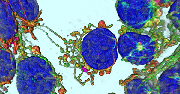 Imaging mitochondria is one of my favorite techniques in the lab. I can stain mitochondria with different fluorescent dyes and visualize the mitochondria in 3D. It's amazing to see and quantify changes happening to organelles only 100 nanometers in size! In the photo to the left, the cobalt blue large ovals are the nuclei (see link in previous section for explanation about the mitochondria structure). The other colors (red, green, magenta) are each staining a different compartment of the mitochondria. The red is a dye which healthy mitochondria can import and is held in the matrix. The green is a fluorescently labeled antibody to TOM20, a protein on the Outer Mitochondrial Membrane that is part of a protein import complex. The magenta is a fluorescently labeled antibody to NME6, another matrix localized protein that is involved in mitochondrial gene expression. The intensities, localization and overlap of the colors help us visualize the distribution of proteins within mitochondria and compare them to other cell types.
I really enjoy sharing best practices for experimental design and execution. Getting good results feels even better when you've designed the experiment with all the appropriate controls and executed it flawlessly. Working as a bench scientist allows me to do creative handiwork every day. It is gratifying to work carefully and rewarding to instill that in others.